
Research
Eye Center Translational Retinal Research Laboratory – Yiu Lab
Retinal diseases such as age-related macular degeneration (AMD) and diabetic retinopathy (DR) are among the major causes of vision loss and blindness in humans. Our laboratory employs a combination of retinal biology and ocular imaging technologies across various animal models including mice, monkeys, and human patients. By developing optically activated drug delivery systems, optogenetic disease models, and gene editing strategies, we hope to better understand the pathophysiology of retinal disorders and discover novel therapies for our patients.
As a clinician-scientist, Glenn Yiu, M.D., Ph.D., spends part of his time providing medical and surgical treatment for patients with retinal diseases, and the rest of his efforts leading our innovative translational research program. Our lab is a multidisciplinary group that collaborates with retinal biologists, biomedical engineers, chemists, veterinarians, and physicians across the UC Davis campus and at other institutions, utilizing ophthalmic imaging, nanotechnology, and genome engineering as our tools. Yiu has received research support from the National Eye Institute, the E. Matilda Ziegler Foundation, Alcon Research Institute, and the ARVO Foundation.
Optogenetic Disease Modeling
The eye is an organ with an inherent optical system. By exploiting this optical system and expressing light-sensitive molecules that can control cellular processes, we are developing systems to optically control biological processes in the retina. For example, we are employing reactive oxygen species (ROS)-generating proteins to allow optogenetic control of oxidative stress in retinal cells as a model of AMD. Further applications of this technology include optical tuning of gene expression using light-sensitive transcription factors.
CRISPR-based Gene Editing
Current treatment of exudative retinal diseases such as neovascular or “wet” age-related macular degeneration (AMD) and diabetic retinopathy includes intraocular injections of drugs that target vascular endothelial growth factor (VEGF). These anti-VEGF agents require frequent injections into the eye, which is costly and a burden to patients. Our laboratory demonstrated the use of novel CRISPR-Cas9 gene editing technology to permanently suppress VEGF secretion from human retinal cells. Using viral vectors that have been used safely in human gene therapy clinical trials, this technology has the potential to permanently suppress aberrant vessel growth (termed “angiogenesis”) in these retinal conditions.
Imaging in Retinal Diseases
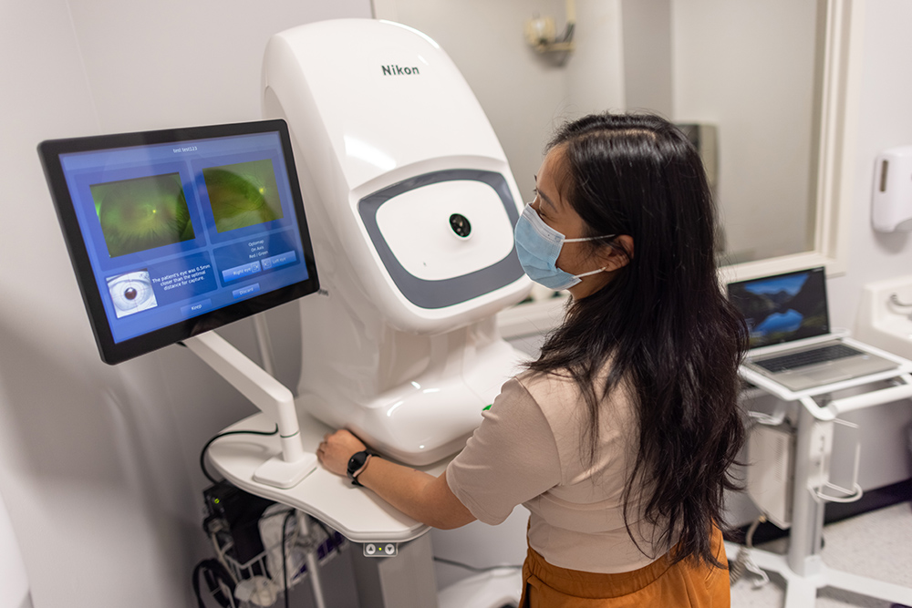
Our ocular imaging research program employs a variety of ocular imaging technologies including optical coherence tomography (OCT) and scanning laser ophthalmoscopy (SLO) across multiple species including mice, nonhuman primates, and human patients. Our past research characterized the so-called suprachoroidal space (a potential space between the choroid and sclera) which may be involved in retinal disease pathogenesis, and possibly a novel route for drug delivery. We are also currently exploring the role of choroidal anatomy in retinal conditions, use of intraoperative OCT in retinal surgery, and utility of imaging biomarkers as prognostic markers. The UC Davis Reading Center also provides comprehensive services for analyzing ophthalmic images from clinical trials.
Primate Research in Macular Degeneration
We collaborate with the California National Primate Research Center to study AMD-like lesions in geriatric rhesus macaques. Because only primates have macula like humans, macular diseases such as AMD are better modeled in nonhuman primates than in common laboratory rodents. Our work in rhesus monkeys allows us to better understand AMD pathogenesis and to test pre-clinical translational therapies that may be readily adapted to human trials.
UC Davis Reading Center
The UC Davis Retinal Reading Center provide comprehensive services for interpreting and analyzing data or images from optical coherence tomography (OCT), color fundus photographs (CFP), fluorescein angiography (FA), infrared reflective (IR), fundus autofluorescence (FAF) including quantitative autofluorescence (qAF), and OCT angiography (OCT-A) from various platforms. Our services support clinical research in various sectors of ophthalmology including retina, glaucoma, cornea, and neuro-ophthalmic studies. Our reading center ensures rigorous quality control, use of validated and reproducible measures, secure data storage and transfer, as well as training, certification, and feedback for ophthalmic photographers and technicians. The Reading Center was under the management of Dr. John L. Keltner, MD and John S. Werner, PhD. Today, Dr. Glenn Yiu serves as the director of the UC Davis Reading Center.
- Ocular Hypertension Treatment Study (OHTS) - The largest glaucoma clinical trial to date that evaluated ocular hypertension and open-angle glaucoma progression in 1636 individuals, with nearly 70,000 visual fields analyzed.
- Optic Neuritis Treatment Trial (ONTT) - This randomized study of 448 patients assessed the role of oral steroids and neuroimaging in the management of optic neuritis. Since 1988, UC Davis has served as the reading center for the 15-year follow-up to this trial and evaluated 18,000 visual fields.
- SHORE Study: We analyzed OCT images from the phase 4, randomized, open-label study comparing intravitreal ranibizumab regimens for treatment of retinal vein occlusions with macular edema.
- TANZANITE Study: We evaluated choroidal and suprachoroidal changes in a phase 2 randomized trial of suprachoroidal triamcinolone and intravitreal aflibercept for macular edema from retinal vein occlusions.
- METforMIN Study: We measured geographic atrophy area in a randomized trial for an oral medication for treatment of end-stage nonexudative age-related macular degeneration.
- PREVENT Study: We analyzed CFP, FA, and FAF images for a prospective, randomized study evaluating prophylactic ranibizumab for prevention of choroidal neovascularization in non-neovascular fellow eyes of patients with unilateral neovascular AMD.
Yiu G, Welch RJ, Wang Y, Wang Z, Wang PW, Haskova Z. Spectral-Domain OCT Predictors of Visual Outcomes after Ranibizumab Treatment for Macular Edema Resulting from Retinal Vein Occlusion. Ophthalmol Retina. 2020 Jan;4(1):67-76. doi: 10.1016/j.oret.2019.08.009. Epub 2019 Aug 28. PubMed PMID: 31669329; PubMed Central PMCID: PMC6944743.
Willoughby AS, Vuong VS, Cunefare D, Farsiu S, Noronha G, Danis RP, Yiu G. Choroidal Changes After Suprachoroidal Injection of Triamcinolone Acetonide in Eyes With Macular Edema Secondary to Retinal Vein Occlusion. Am J Ophthalmol. 2018 Feb;186:144-151. doi: 10.1016/j.ajo.2017.11.020. Epub 2017 Dec 2. PubMed PMID: 29199012; PubMed Central PMCID: PMC5805638.
Keltner JL, Johnson CA, Cello KE, Bandermann SE, Fan J, Levine RA, Kass MA, Gordon MO. Visual field quality control in the Ocular Hypertension Treatment Study (OHTS). J Glaucoma. 2007 Dec;16(8):665-9. doi: 10.1097/IJG.0b013e318057526d. PubMed PMID: 18091452.
Beck RW, Trobe JD, Moke PS, Gal RL, Xing D, Bhatti MT, Brodsky MC, Buckley EG, Chrousos GA, Corbett J, Eggenberger E, Goodwin JA, Katz B, Kaufman DI, Keltner JL, Kupersmith MJ, Miller NR, Nazarian S, Orengo-Nania S, Savino PJ, Shults WT, Smith CH, Wall M. High- and low-risk profiles for the development of multiple sclerosis within 10 years after optic neuritis: experience of the optic neuritis treatment trial. Arch Ophthalmol. 2003 Jul;121(7):944-9. doi: 10.1001/archopht.121.7.944. PubMed PMID: 12860795.
Johnson CA, Keltner JL, Cello KE, Edwards M, Kass MA, Gordon MO, Budenz DL, Gaasterland DE, Werner E. Baseline visual field characteristics in the ocular hypertension treatment study. Ophthalmology. 2002 Mar;109(3):432-7. doi: 10.1016/s0161-6420(01)00948-4. PubMed PMID: 11874743.
Keltner JL, Johnson CA, Beck RW, Cleary PA, Spurr JO. Quality control functions of the Visual Field Reading Center (VFRC) for the Optic Neuritis Treatment Trial (ONTT). Control Clin Trials. 1993 Apr;14(2):143-59. doi: 10.1016/0197-2456(93)90016-7. PubMed PMID: 8500303.